NewsPhotography

Olympus Unveils the Winners of the Best Life Science Photos of 2020

Olympus has announced the winners of its Global Image of the Year Life Science Light Microscopy Award, which is an annual competition that recognizes the best in life science photos taken with a microscope.

This competition is hosted by Olympus’s Life Sciences division, which is not to be confused with the newly-formed OM Digital under Japan Industrial Partners. The competition started in 2017 as the Image of the Year European Life Science Light Microscopy Award with the aim to celebrate both the artistic and scientific value of microscopy images. Today, the organizers say that the competition stays true to this mission by encouraging people across the world to look at scientific images in a new way, appreciate their beauty, and share images with others.

To be considered for this year’s competition, images had to be submitted between September 15, 2020 and January 31, 2021. A maximum of three images was allowed per participant, and anyone over the age of 18 could participate (provided that they are not an Olympus employee, family member, judge, or family member of a judge nor engaged in the fields of manufacturing or sales of microscopes).

All entries were evaluated on artistic and visual aspects, scientific impact, and microscope proficiency.

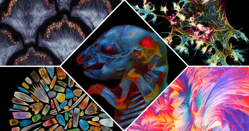
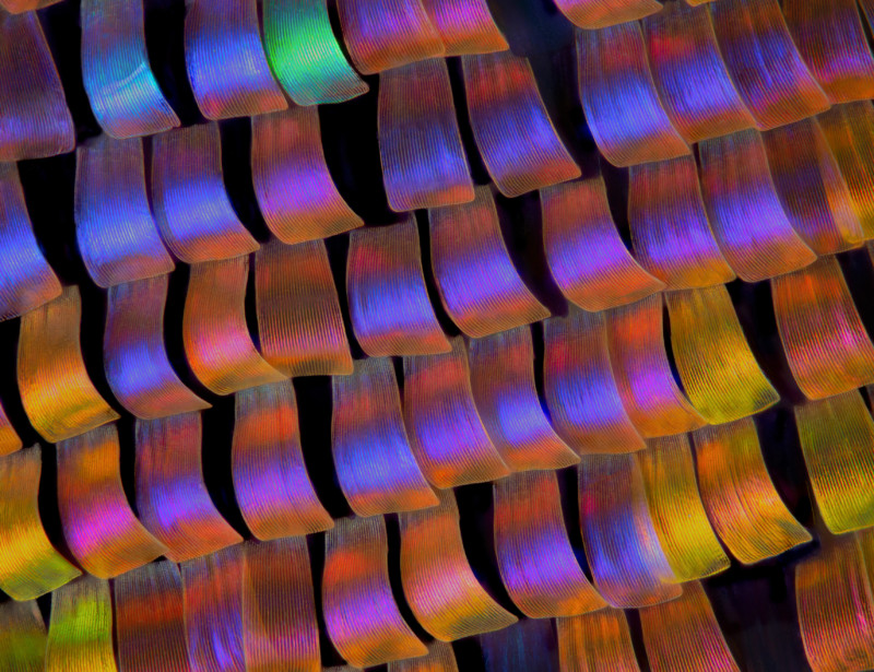
From rat embryos to butterfly scales and snakeskin, the Olympus Life Science team was impressed by the diverse collection captured under the microscope this year.

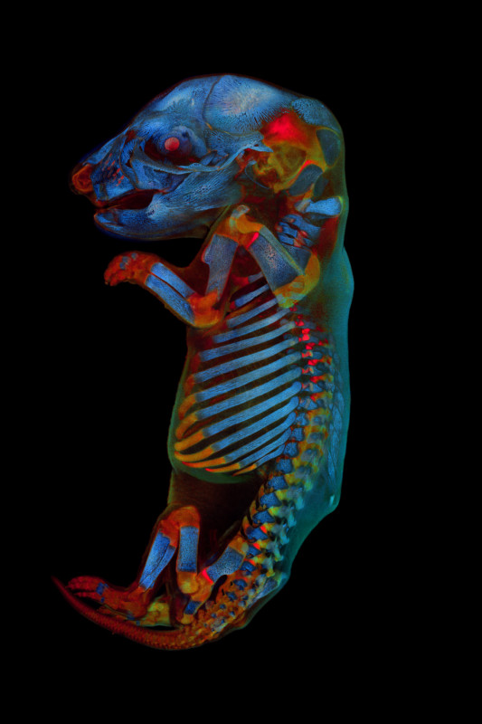
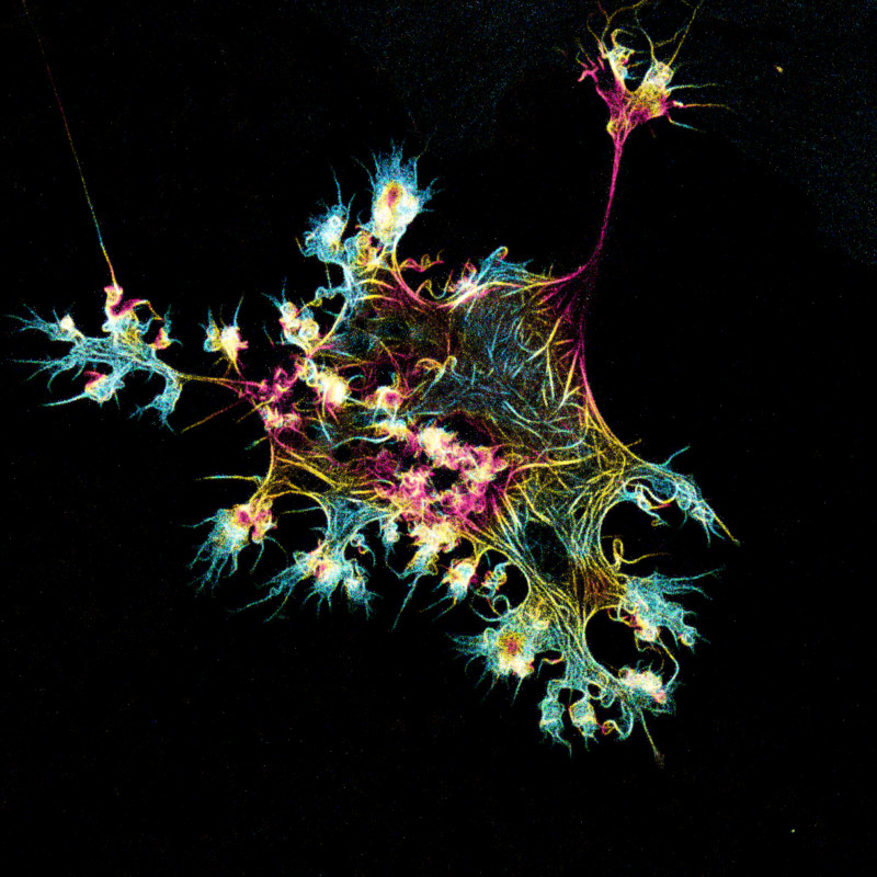
This year’s competition saw nearly 700 submissions from 61 different countries, with the winning image going to Werner Zuschratter from Germany. Zuschratter submitted the incredible photo below depicting a whole rat embryo that he captured with a confocal microscope. For the grand prize, Zuschratter will receive an Olympus SZX7 stereo microscope with a DP27 digital camera.

In addition to the overall global winner, three regional awards were presented to XinPei Zhang (China) for Asia, Justin Zoll (USA) for the Americas, and Grigorii Timin (Switzerland) for EMEA. Each regional winner will receive an Olympus CX23 upright microscope.

Collagen fibers (second harmonic generation) and dermal pigment cells (autofluorescence) in African house snake embryonic skin; maximal intensity projection of 10 confocal slices. | Regional Winner – EMEA – Grigorii Timin – Switzerland
Scales collected from the wings of over 40 species of butterflies were photographed individually and finally assembled into this image. | Regional Winner – Asia Pacific – XinPei Zhang – China
Polarized light microscopy panorama of L-glutamine and beta alanine crystals | Regional Winner – Americas – Justin Zoll – United States

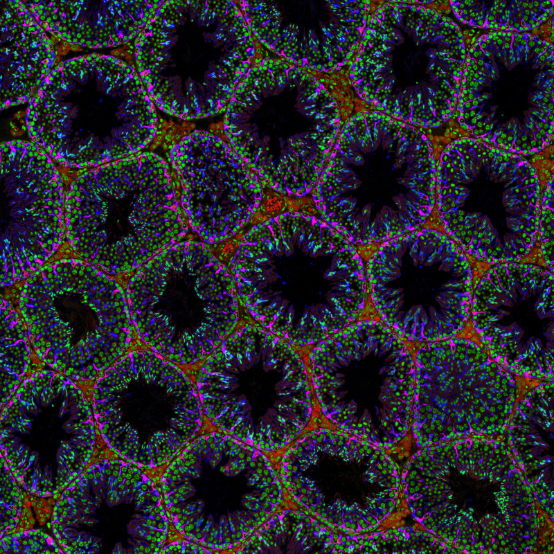
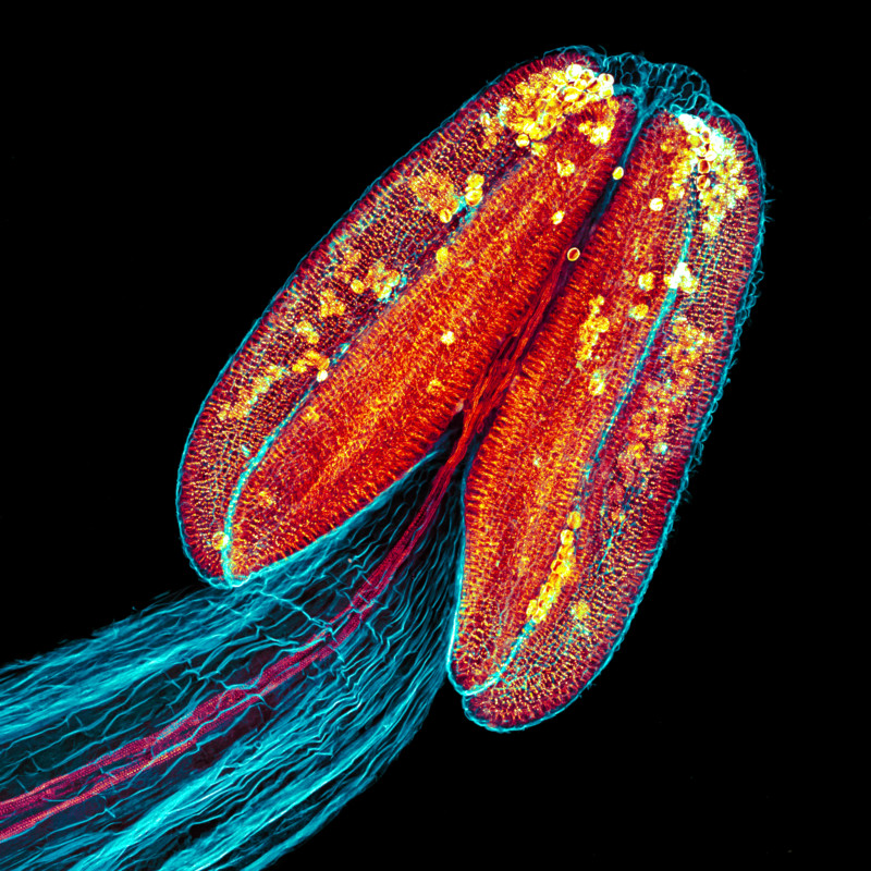
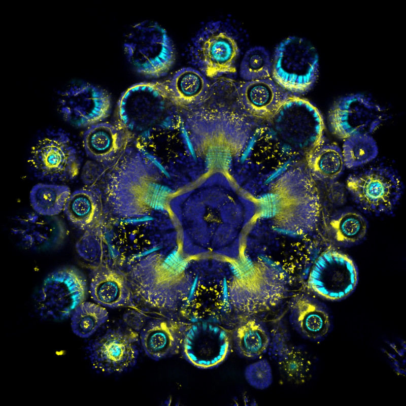
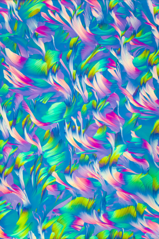
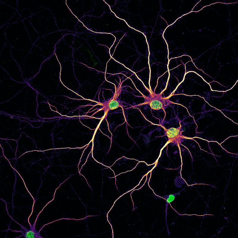
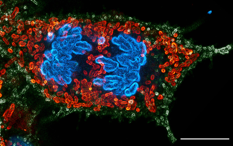
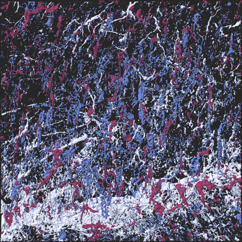
Though they may not have brought home the grand prize or won in any of the three regions, the honorable mentions below are still breathtaking images.

Mouse testes stained for Sertoli cells (blue and red) | Derek Sung, United States
Anther of Arabidopsis arenosa stained with aniline blue, Z-stack of confocal sections (maximum intensity projection) | Jan Martinek, Czech Republic
Whole-mount immunofluorescence showing the muscles (cyan, F-actin) and the nervous system (yellows, acetylated tubulin) counterstained by the nuclei (blue, DNA) of a clarified and decalcified post-metamorphic sea urchin juvenile (Paracentrotus lividus). | Laurent Formery, France
A polarized light micrograph of crystals formed by the rapid evaporation of water from an aqueous solution of Beta-Alanine (a popular training supplement), and L-glutamine (an essential amino acid). Home-made tunable wave plate. | Matt Inman, Australia
Microfilament structure in U2OS labeled with fluorescent protein, 3D projection image acquired by airyscan | MingShu Zhang, China
Primary rat cortical neurons at 14 days in vitro with nuclei (green) and developing dendrites (mpl-inferno LUT) | Nadia Efimova, United States
The sample shows a dividing human embryonic kidney (HEK) 293 cell. Green marks the cellular boundary, red marks the mitochondria, while blue shows the separating chromosomes. | Sayantan Datta, India
Wing scales of the twilight moth Urania ripheus. | Walter Ferrari, Argentina
“Pollock’s glia”. 3D reconstructed immunostaining image of astrocyte (GFAP+, white), oligodendrocyte (NG2+, blue), and microglia (IBA1+, red) in the brain white matter, which is highly similar to Pollock’s Abstract paintings. | YiXun Su, China

To read about the jurors and see past competition winners, visit the Olympus Lifescience website here.


Image credits: All photos individually credited and provided courtesy of Olympus Life Science.


Author: Jaron Schneider
Source: Petapixel

Related posts
AI & RoboticsNews

Hacking internal AI chatbots with ASCII art is a security team’s worst nightmare

AI & RoboticsNews

Microsoft launches new Azure AI tools to cut out LLM safety and reliability risks

AI & RoboticsNews

AI21 Labs juices up gen AI transformers with Jamba

DefenseNews

Northrop says Air Force design changes drove higher Sentinel ICBM cost

Sign up for our Newsletter and
stay informed!